
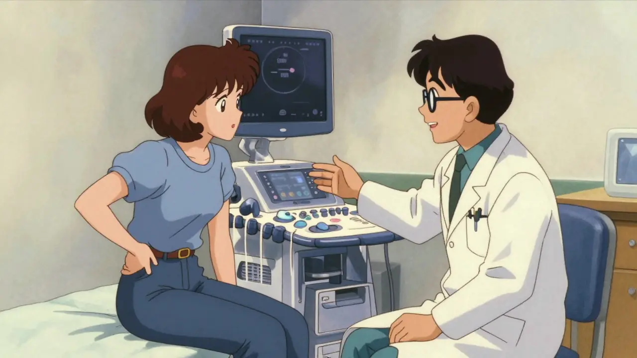
Imagine feeling a sharp pain in your side or lower back. Your doctor suspects a blockage in your urinary tract, but they don't want to expose you to radiation. This is where renal ultrasound comes in. It is the go-to test for checking kidney health because it uses sound waves instead of X-rays. You get clear images of your kidneys, bladder, and ureters without any needles or contrast dyes. It’s fast, safe, and gives immediate answers about whether urine is backing up into your kidneys.
This guide breaks down exactly what happens during the test, what normal numbers look like, and how doctors use these results to diagnose conditions like kidney stones or congenital blockages. We’ll also look at why this test beats CT scans for many patients, especially children and pregnant women.
Why Renal Ultrasound Is the First Choice
When doctors suspect a problem with your kidneys, they need a tool that shows structure clearly. Renal ultrasound is a non-invasive imaging technique that uses high-frequency sound waves to visualize kidney anatomy. It has been the standard first-line diagnostic tool since the 1970s. Why? Because it avoids ionizing radiation entirely. A CT scan, which is often used for kidney stones, delivers about 10 millisieverts (mSv) of radiation. An ultrasound delivers zero. This makes it the safest option for repeated monitoring.
The cost is another factor. In the United States, a renal ultrasound typically costs between $200 and $500. Compare that to Magnetic Resonance Urography (MRU), which can run $1,500 to $2,500, or even a basic CT urography. For most initial evaluations, ultrasound provides enough detail to decide the next steps without breaking the bank. According to the American College of Radiology, about 12 million renal ultrasounds are performed annually in the US alone, proving its reliability and accessibility.
However, it isn’t perfect. If you have a very small stone-say, 1 or 2 millimeters-ultrasound might miss it. CT scans detect stones as small as 1 mm with nearly 100% accuracy. Ultrasound catches about 80% of stones larger than 3 mm. But for detecting the *effects* of a blockage, like swelling in the kidney, ultrasound is unmatched in safety and speed.
What Doctors Measure: Size and Structure
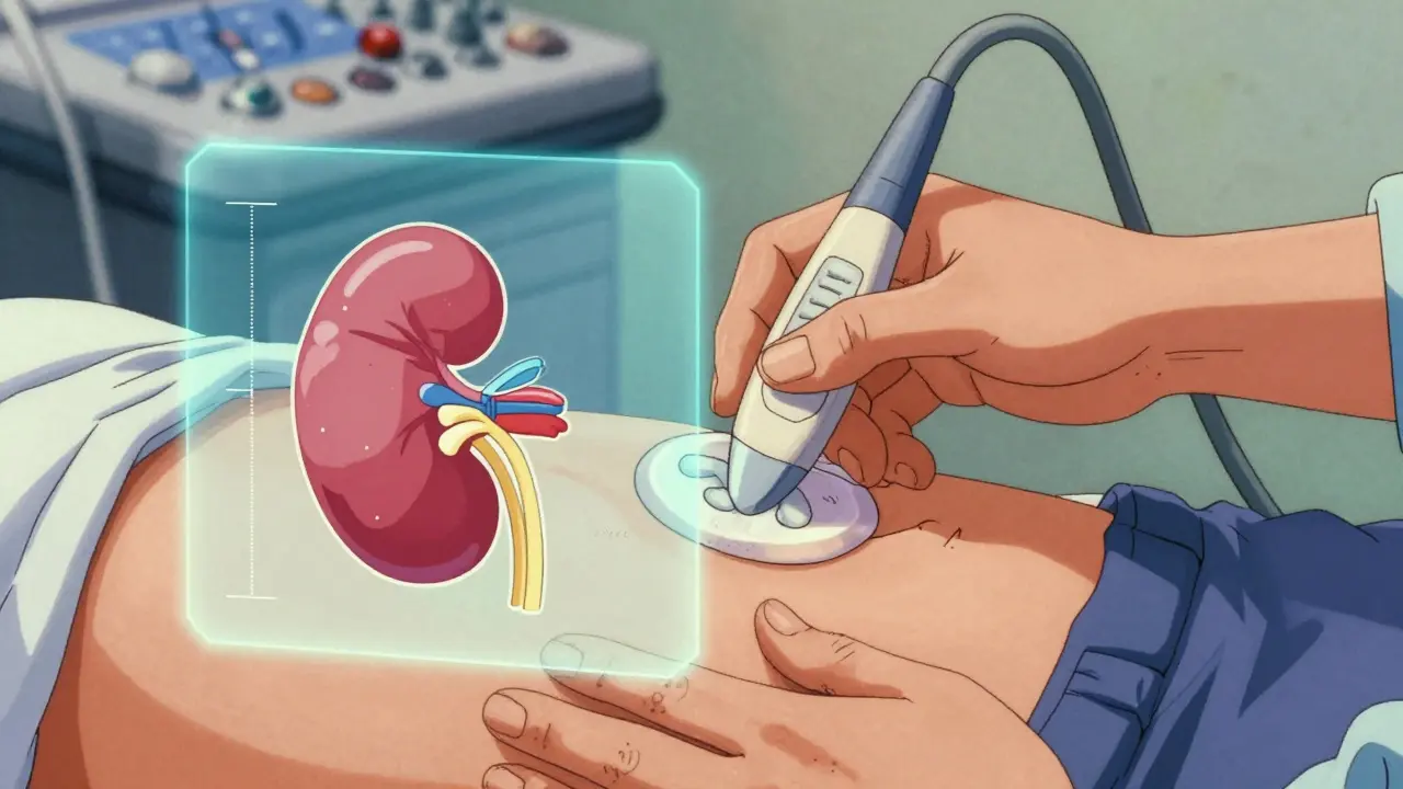
During the exam, the sonographer places a probe on your abdomen and back. They aren’t just looking for lumps; they are measuring specific dimensions to assess kidney health. Here are the key metrics they track:
- Kidney Length: Normal adult kidneys measure between 9 and 13 centimeters. If a kidney is significantly smaller, it might indicate chronic damage or shrinkage from long-term disease. If it’s larger, it could signal acute inflammation or obstruction.
- Cortical Thickness: The cortex is the outer layer of the kidney where filtration happens. Healthy tissue should be thicker than 1 centimeter. Thinning cortex suggests chronic kidney disease or long-standing pressure from obstruction.
- Renal Pelvis Diameter: This is the central collecting area for urine. Normally, the anteroposterior diameter is less than 7 millimeters. If it’s wider, urine is pooling, which is a sign of blockage.
These measurements give a snapshot of the kidney’s physical state. But size alone doesn’t tell the whole story. A kidney can be large but functioning poorly, or small but still filtering well. That’s why doctors combine these structural measurements with functional tests, like blood work or Doppler flow analysis.
Detecting Obstruction: Hydronephrosis and Grading
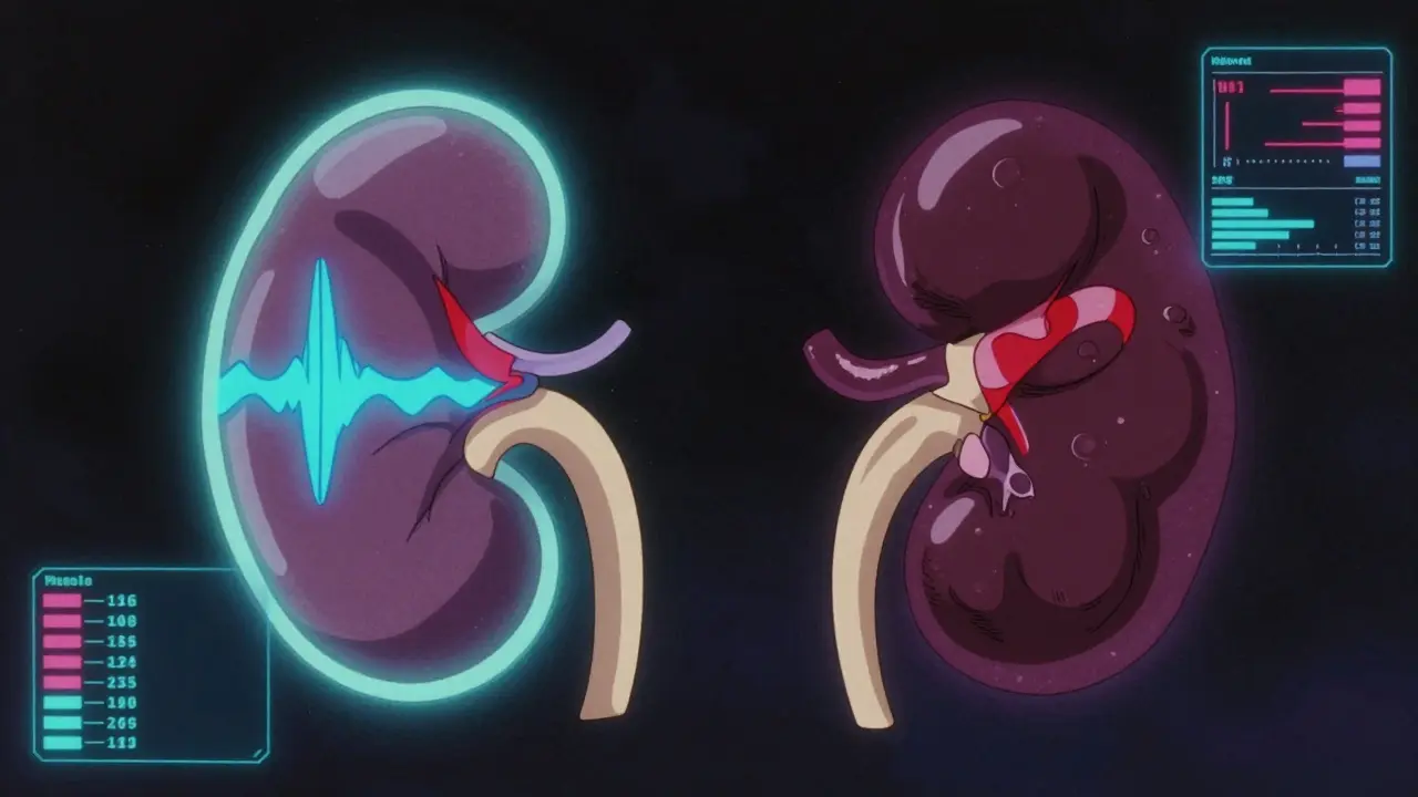
The most common reason for ordering a renal ultrasound is to check for hydronephrosis, which is the dilation of the renal pelvis due to urine accumulation caused by an obstruction. When urine can’t flow freely out of the kidney, it backs up, stretching the internal structures. This pressure can damage kidney tissue over time if not relieved.
Doctors grade hydronephrosis using systems like the Society for Fetal Urology (SFU) scale. It ranges from Grade 0 (normal) to Grade 4 (severe).
- Grade 1: Mild dilation of the renal pelvis only.
- Grade 2: Dilation extends to the calyces (smaller branches).
- Grade 3: Moderate dilation with thinning of the kidney wall.
- Grade 4: Severe dilation with significant cortical thinning, indicating potential permanent damage.
In emergency rooms, point-of-care (POC) ultrasound is often used for patients with suspected renal colic (kidney stone pain). Studies show POC ultrasound has a sensitivity of about 70% and specificity of 75% for detecting stones compared to CT. While not perfect, it’s fast. Emergency physicians report it cuts diagnosis time by roughly 45 minutes. This speed matters when you’re in severe pain and need treatment decisions quickly.
Blood Flow Analysis: The Resistive Index
Structure tells you what the kidney looks like; blood flow tells you how well it’s working under pressure. Using Doppler ultrasound, doctors measure the Resistive Index (RI), a calculation derived from peak systolic and end-diastolic velocities in renal arteries. The formula is simple: (Peak Systolic Velocity - End Diastolic Velocity) / Peak Systolic Velocity.
A normal RI is usually below 0.70. When an obstruction blocks urine flow, pressure builds up inside the kidney. This pressure squeezes the tiny blood vessels, increasing resistance to blood flow. Consequently, the RI rises. A study published in the Nigerian Journal of Clinical Practice found that an RI of ≥0.70 has an 86.7% sensitivity and 90% specificity for diagnosing obstructive uropathy. This means if your RI is high, there’s a strong chance an obstruction is causing increased pressure.
This metric is crucial for distinguishing between a simple, temporary backup and a serious blockage that needs intervention. It helps doctors decide if you need a stent or surgery to relieve the pressure before permanent damage occurs.
| Feature | Renal Ultrasound | CT Urography | MRI/MRU |
|---|---|---|---|
| Radiation Exposure | None | High (~10 mSv) | None |
| Stone Detection | Good (>3mm stones) | Excellent (all sizes) | Poor |
| Soft Tissue Detail | Moderate | High | Very High |
| Cost (USD) | $200 - $500 | $1,000 - $2,500 | $1,500 - $2,500 |
| Best For | Initial screening, pregnancy, kids | Complex stones, trauma | Tumors, vascular issues |
Limitations and When Other Tests Are Needed
Ultrasound is great, but it has blind spots. The biggest limitation is body habitus. Sound waves struggle to penetrate thick layers of fat. If a patient has a BMI over 35, the image quality drops significantly. Radiologists often note that in obese patients, ultrasound becomes unreliable, forcing them to order a CT scan instead. Bowel gas is another enemy; air blocks sound waves, sometimes hiding parts of the kidney or ureter.
Another gap is functional data. Traditional ultrasound shows anatomy, not function. It can’t tell you exactly how much urine is draining per minute. For that, doctors might order a diuretic renogram, a nuclear medicine test that tracks radioactive tracer through the kidneys. While it involves radiation, it provides precise drainage rates that ultrasound cannot match.
Also, ultrasound is operator-dependent. A skilled sonographer gets better images than a novice. Studies show up to 20% variation in kidney length measurements between experienced and inexperienced operators. This is why accreditation matters. The American Institute of Ultrasound in Medicine (AIUM) recommends at least 40 supervised exams for certification to ensure consistent quality.
New Technologies: Elastography and AI
The field isn’t standing still. New techniques are making ultrasound more powerful. One exciting development is shear-wave elastography (SWE). This technology measures the stiffness of kidney tissue. As obstruction pressure increases, kidney tissue becomes stiffer. Research by Gennisson et al. showed a linear increase in stiffness related to urinary pressure. This could allow doctors to quantify obstruction severity without waiting for changes in size or blood flow.
Artificial Intelligence (AI) is also entering the room. Mayo Clinic and other institutions are testing AI algorithms that automatically grade hydronephrosis. These tools reduce human error and speed up reporting. Imagine an AI that instantly flags a dangerous level of swelling while the sonographer is still scanning. This integration promises to make ultrasound even more reliable and accessible.
Super-resolution ultrasound imaging is another frontier. It allows visualization of microvascular changes in the kidney’s cortex. This could help detect early signs of fibrosis or scarring before they show up on standard scans. These advancements suggest that ultrasound will remain the dominant first-line tool for decades to come, evolving from a static image provider to a dynamic, quantitative diagnostic powerhouse.
Do I need to prepare for a renal ultrasound?
Generally, no special preparation is needed. However, some doctors may ask you to drink water beforehand to fill your bladder. A full bladder acts as an acoustic window, pushing bowel loops aside and providing a clearer view of the kidneys and ureters. Follow your specific doctor’s instructions regarding fasting or hydration.
Is renal ultrasound painful?
No, the procedure is completely painless. The sonographer applies gel to your skin and moves a transducer (probe) over your abdomen and back. You might feel slight pressure, but there is no pain, needles, or radiation involved.
Can ultrasound detect all kidney stones?
Not all of them. Ultrasound is excellent for finding larger stones (greater than 3-4 mm) and those in the kidney or bladder. However, it often misses small stones in the ureter (the tube connecting kidney to bladder) due to interference from bowel gas. If suspicion remains high after a negative ultrasound, a CT scan is usually the next step.
What does a high Resistive Index mean?
A high Resistive Index (typically ≥0.70) indicates increased resistance to blood flow within the kidney. This is often caused by pressure from an obstruction, such as a stone or stricture, blocking urine flow. It can also be seen in severe kidney infections or vascular diseases. Your doctor will correlate this number with your symptoms and other findings.
How accurate is ultrasound for kidney size?
Ultrasound is highly accurate for measuring kidney length, with a normal range of 9-13 cm for adults. However, accuracy depends on the skill of the sonographer. Experienced technicians minimize measurement errors. Variations of up to 20% can occur between novice and expert operators, so consistency in who performs your follow-up scans is beneficial.



kavitha kumar - 8 July 2026
so basically if u r fat they cant see anything lol
i had this done last year and the tech was like oh well we will try our best but ur body habitus is not ideal
it was so rude she said it right in front of my husband
and then i had to pay for a ct scan anyway which cost me like $2000 out of pocket because insurance denied it first time
why do doctors always assume everyone is skinny???
fred morgan - 10 July 2026
The distinction between structural imaging and functional assessment is critical here. While renal ultrasound provides excellent anatomical detail regarding size and obstruction, it lacks the capability to quantify glomerular filtration rate or precise drainage dynamics. Consequently, reliance on ultrasound alone for complex cases may lead to diagnostic delays. The resistive index mentioned is a useful adjunct, yet it remains operator-dependent and susceptible to systemic vascular influences unrelated to local obstruction. Patients should understand that a normal ultrasound does not definitively rule out pathology, particularly in the context of persistent symptoms.
Sarah Boomgaard - 10 July 2026
you people think technology fixes everything.
it does not.
the human body is not a machine to be scanned and fixed with waves.
we have lost touch with the simple truth that pain is a signal from nature.
instead of listening to our bodies we run to machines that tell us numbers.
a high resistive index means nothing if the spirit is broken.
this obsession with measuring millimeters is a symptom of a sick society that fears uncertainty.
let the kidney heal itself by stopping the stress and eating real food not processed garbage.
ultrasound is just a distraction from the real problem which is modern life.
Gilbert Bankual - 11 July 2026
Great info on the SFU grading scale. Knowing what Grade 1 vs Grade 4 looks like helps patients advocate for themselves. If you have severe dilation, do not wait. Get that stent or surgery before permanent damage sets in. Early detection saves kidneys. Keep pushing for better access to these safe tests.
Ashley Loera - 13 July 2026
i hate how they say its painless but the gel is so cold it hurts
and then the probe pushes hard on your back and you cant breathe
also why do they always make you turn around so much?
my lower back was sore for days after
not to mention the waiting room smell
but yeah whatever it saves money i guess
just wish they would warn you about the pressure part
Dawn Renee - 13 July 2026
of course the big pharma companies want you to believe ultrasound is enough so they dont sell you more expensive scans
its a conspiracy to keep healthcare costs low while hiding the truth about small stones
they know CT scans find the tiny ones but they push ultrasound first to delay diagnosis
think about it why would they give you the best test immediately?
they want you to suffer longer so you need more treatment later
do not trust the radiologists they are paid off by the equipment manufacturers
get a second opinion always question the results
Divya Prakash - 15 July 2026
It is truly fascinating how the layperson continues to underestimate the profound technical nuances inherent in shear-wave elastography, a modality that represents the vanguard of non-invasive tissue characterization, thereby rendering obsolete the archaic reliance on mere morphological dimensions which, as any competent sonographer will attest, are fraught with inter-observer variability that plagues the field of diagnostic radiology to such an extent that one might wonder if the American Institute of Ultrasound in Medicine has abdicated its duty to enforce rigorous standards of competence among its practitioners who often fail to appreciate the subtle gradations of stiffness that correlate linearly with intrarenal pressure, thus necessitating a deeper understanding of the biophysical principles governing acoustic wave propagation in heterogeneous media rather than simply relying on the superficial allure of artificial intelligence algorithms which, while promising, lack the nuanced interpretive framework required to distinguish between transient physiological changes and pathological fibrosis in the renal cortex.